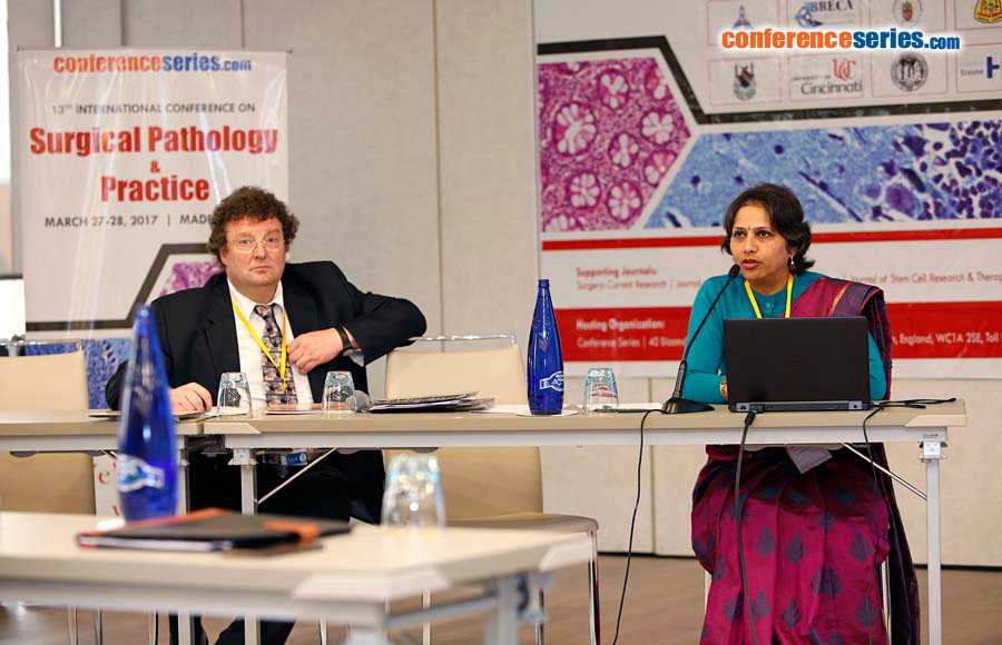
Uma Nahar Saikia
Postgraduate Institute of Medical Education and Research, India
Title: Histological spectrum of pure neuritic leprosy: A tertiary centre experience
Biography
Biography: Uma Nahar Saikia
Abstract
Introduction: Pure neuritic leprosy (PNL) is a rare presentation of leprosy and defined isolated involvement of one or more nerves by leprosy in absence of skin involvement.
Aim: The aim of this study is to assess the histological spectrum of PNL.
Materials & Methods: A retrospective analysis of all histological proven PNL cases (20) from time period June 2000 to 2016 was done. Detailed histopathological examination of nerve biopsy was performed. Myelin and axonal status were evaluated by using myelin stain and immunohistochemistry for neurofilament protein (NFP) respectively.
Results: The suspected clinical diagnosis was Hansen in 82.3%, Mononeuritis multiplex in 11.7% and demyelination in 5.8% cases. Moderate to marked perineural thickening was seen in 70.5% cases. Moderate to severe perineural and endoneureal inflammation was seen in 75% and 95% of cases respectively. Granuloma formation was seen both in endoneureal (75%) and perineureal (15%) location. Foam cell infiltration was more common in endoneureal (65%) than perineureal (20%) location. Necrosis (5%) and vasculitis (10%) were seen occasionally. Severe myelin and axonal loss was seen in 65%. Lepra bacilli were positive in 10 cases. One case had normal morphology with lepra bacilli positivity. There was no significant histological difference between lepra bacilli positive and negative cases.
Conclusion: Leprosy is known to cause endoneureal inflammation, perineural thickening, inflammation or even granulomas in PNL. Necrosis and vasculitis are rarely noted. Myelin and axonal loss are almost universal. Even if morphologically biopsy is normal, lepra stain should be performed on all the suspected PNL cases.


