Day 2 :
Keynote Forum
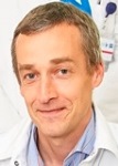
Jürgen Hescheler
University of Cologne, Germany
Keynote: Pluripotent stem cells for research and clinical application
Time : 10:00-10:40

Biography:
Jürgen Karl-Josef Hescheler (born 2 May 1959) is a German physician and stem cell researcher. He is director to the Institute for Neurophysiology and a university professor at the University of Cologne. He is one of the most eminent and the most productive stem cell researcher’s worldwide, averaging more than 1000 citations of his publications per year. He has been working with embryonic stem cells since the late 80’s. He became the first researcher to accomplish an electrophysiological characterisation of stem cells and was also among the first scientists in Germany obtaining permission to do research on human embryonic stem cells.
Abstract:
There is no doubt that work on stem cells will be the most promising approach for the medicine of the 21st century and probably revolutionize the therapy of many diseases including cardiac infarction and failure, diabetes, Parkinson’s disease, spinal cord lesion etc. It is our aim to provide a fundamental basis to the development of new medical treatments. Stem cell research is a broad field which requires nearly all techniques of modern life science such as genetics, cell biology, physiology, biochemistry, histology, etc., but it also requires the input of experimental surgery and bioengineering technologies. Induced pluripotent stem (iPS) cells represent the most promising approach for future stem cell-based tissue repair in regenerative medicine. iPS cells are functionally highly similar to embryonic stem (ES) cells, but have in addition the advantage of being ethically non-controversial, and are obtainable from readily accessible autologous sources. However, although proof of principle for the therapeutic use of iPS cells in neuronal and cardiac diseases has been shown both at the laboratory scale and in animal models, the methods used today for generation, cultivation, differentiation and selection still have to be translated for their later clinical usage. This presentation will give an overview on our recent research work on human embryonic in comparison with iPS cells. Starting from our basic investigations on the physiological properties of cardiomyocytes developed from pluripotent stem cells we have established in vitro and in vivo transplantation models enabling us to systematically investigate and optimize the physiological integration and regeneration of the diseased tissue. Our main focus is the cardiac infarction model. Moreover, in vitro culture and expansion of stem cells is far from optimal and needs further research in order to overcome problems related to insufficient numbers of obtained stem cells and aging of the obtained stem cell population.
Keynote Forum
Shahla Masood
University of Florida College of Medicine, USA
Keynote: Borderline breast disease: An entity to minimize errors in over diagnosis of low grade ductal carcinoma in situ in breast pathology
Time : 10:40-11:20

Biography:
Shahla Masood is currently a Professor and Chair of the Department of Pathology at University of Florida College of Medicine, Jacksonville and Chief of Pathology and Laboratory Medicine at Shands Jacksonville. She is also the Director of the Pathology Residency Training Program, as well as Cytopathology and Breast Pathology Fellowship Training Program. In addition, she is the Medical Director of Shands Jacksonville Breast Health Center. An internationally recognized expert in breast cancer diagnosis and prognosis, she has fostered the concept of an integrated multidisciplinary approach in breast cancer care, research and education. She has recently been appointed to chair a committee of the National Accreditation Program for Breast Centers (NAPBC) with a new initiative to explore the possibility of expansion of this program to an international level.
Abstract:
During the last several years, increased public awareness, advances in breast imaging and enhanced screening programs have led to early breast cancer detection and attention to cancer prevention. The numbers of image-detected biopsies have increased and pathologists are expected to provide more information with smaller tissue samples. These biopsies have resulted in detection of increasing numbers of high-risk proliferative breast diseases and in situ cancers. The general hypothesis is that some forms of breast cancers may arise from established forms of ductal carcinoma in situ (DCIS) and atypical ductal hyperplasia (ADH) and possibly from more common forms of ductal hyperplasia. However, this is an oversimplification of a very complex process, given the fact that the majority of breast cancers appears to arise de-novo or from a yet unknown precursor lesion. Currently, ADH and DCIS are considered as morphologic risk factors and precursor lesions for breast cancer. However, morphologic distinction between these two entities has remained a real issue that continues to lead to over diagnosis and over treatment. Aside from morphologic similarities between ADH and low grade DCIS, biomarker studies and molecular genetic testing’s have shown that morphologic overlaps are reflected at the molecular levels and raise questions about the validity of separating these two entities. It is hoped that as we better understand the genetic basis of these entities in relation to ultimate patient outcome, the suggested use of the term of “Borderline Breast Disease” can minimize the number of patients who are subject to overtreatment.
- Surgical Pathology and Diagnosis | Surgical Pathology Practice Issues and Concerns | Cancer Pathology | Breast Pathology | Gynecologic Pathology | Neuro Surgery and Pathology | Gastrointestinal and Liver Pathology | Dermatopathology | Genitourinary Pathology | Histologic Pathology | Bone and Soft Tissue Pathology
Session Introduction
Jürgen Hescheler
Institute for Neurophysiology- University of Cologne, Germany
Title: Pluripotent stem cells for research and clinical application

Biography:
Jürgen Karl-Josef Hescheler (born 2 May 1959) is a German physician and stem cell researcher. He is director to the Institute for Neurophysiology and a university professor at the University of Cologne. He is one of the most eminent and the most productive stem cell researcher’s worldwide, averaging more than 1000 citations of his publications per year. He has been working with embryonic stem cells since the late 80’s. He became the first researcher to accomplish an electrophysiological characterisation of stem cells and was also among the first scientists in Germany obtaining permission to do research on human embryonic stem cells.
Abstract:
There is no doubt that work on stem cells will be the most promising approach for the medicine of the 21st century and probably revolutionize the therapy of many diseases including cardiac infarction and failure, diabetes, Parkinson’s disease, spinal cord lesion etc. It is our aim to provide a fundamental basis to the development of new medical treatments. Stem cell research is a broad field which requires nearly all techniques of modern life science such as genetics, cell biology, physiology, biochemistry, histology, etc., but it also requires the input of experimental surgery and bioengineering technologies. Induced pluripotent stem (iPS) cells represent the most promising approach for future stem cell-based tissue repair in regenerative medicine. iPS cells are functionally highly similar to embryonic stem (ES) cells, but have in addition the advantage of being ethically non-controversial, and are obtainable from readily accessible autologous sources. However, although proof of principle for the therapeutic use of iPS cells in neuronal and cardiac diseases has been shown both at the laboratory scale and in animal models, the methods used today for generation, cultivation, differentiation and selection still have to be translated for their later clinical usage. This presentation will give an overview on our recent research work on human embryonic in comparison with iPS cells. Starting from our basic investigations on the physiological properties of cardiomyocytes developed from pluripotent stem cells we have established in vitro and in vivo transplantation models enabling us to systematically investigate and optimize the physiological integration and regeneration of the diseased tissue. Our main focus is the cardiac infarction model. Moreover, in vitro culture and expansion of stem cells is far from optimal and needs further research in order to overcome problems related to insufficient numbers of obtained stem cells and aging of the obtained stem cell population.
Shahla Masood
University of Florida College of Medicine, USA
Title: Borderline breast disease: An entity to minimize errors in over diagnosis of low grade ductal carcinoma in situ in breast pathology

Biography:
Shahla Masood is currently a Professor and Chair of the Department of Pathology at University of Florida College of Medicine, Jacksonville and Chief of Pathology and Laboratory Medicine at Shands Jacksonville. She is also the Director of the Pathology Residency Training Program, as well as Cytopathology and Breast Pathology Fellowship Training Program. In addition, she is the Medical Director of Shands Jacksonville Breast Health Center. An internationally recognized expert in breast cancer diagnosis and prognosis, she has fostered the concept of an integrated multidisciplinary approach in breast cancer care, research and education. She has recently been appointed to chair a committee of the National Accreditation Program for Breast Centers (NAPBC) with a new initiative to explore the possibility of expansion of this program to an international level.
Abstract:
During the last several years, increased public awareness, advances in breast imaging and enhanced screening programs have led to early breast cancer detection and attention to cancer prevention. The numbers of image-detected biopsies have increased and pathologists are expected to provide more information with smaller tissue samples. These biopsies have resulted in detection of increasing numbers of high-risk proliferative breast diseases and in situ cancers. The general hypothesis is that some forms of breast cancers may arise from established forms of ductal carcinoma in situ (DCIS) and atypical ductal hyperplasia (ADH) and possibly from more common forms of ductal hyperplasia. However, this is an oversimplification of a very complex process, given the fact that the majority of breast cancers appears to arise de-novo or from a yet unknown precursor lesion. Currently, ADH and DCIS are considered as morphologic risk factors and precursor lesions for breast cancer. However, morphologic distinction between these two entities has remained a real issue that continues to lead to over diagnosis and over treatment. Aside from morphologic similarities between ADH and low grade DCIS, biomarker studies and molecular genetic testing’s have shown that morphologic overlaps are reflected at the molecular levels and raise questions about the validity of separating these two entities. It is hoped that as we better understand the genetic basis of these entities in relation to ultimate patient outcome, the suggested use of the term of “Borderline Breast Disease” can minimize the number of patients who are subject to overtreatment.
Pieter Demetter
Erasme University Hospital, Belgium
Title: Benign, premalignant and malignant pancreatic cystic lesions: The pathology landscape

Biography:
Pieter Demetter has completed his PhD from Ghent University and currently working at the Université Libre de Bruxelles. He is Head of the Clinic for Digestive Pathology at the Pathology Department of the Erasme University Hospital. He has published more than 130 papers in reputed journals, serves as Associate Editor of Histopathology and is Vice-President of the Belgian Society of Pathology.
Abstract:
Pancreatic cystic lesions are being increasingly detected in last years by significant improvement in imaging technologies, increased awareness of their existence and the growth of the aging population. These lesions are a broad group of pancreatic tumours with varying demographical, morphological, clinical and histological characteristics. Pancreatic cysts can be classified grossly into pseudocysts and true cysts. Pseudocysts develop mostly 4 weeks after the onset of acute pancreatitis, and are the natural evolution of acute fluid collections. In the true cysts group, it is important to distinguish mucinous from non-mucinous cysts because the former are considered being premalignant lesions. Distinguishing between the various types of lesions has important prognostic and therapeutic implications. The sensitivity of cytology varies depending on the expertise of the endoscopist and the pathologist. Diagnostic accuracy can increase up to 80-90% if cytology is complemented with measurements of CEA, amylase levels and mucin staining. Although cyst fluid analysis is a tool in preoperative diagnosis, final diagnosis if often obtained only after histopathological analysis. Molecular markers in the cyst fluid are being increasingly studied in recent years. Molecular tests of the aspirated cystic fluid seem particularly useful for detecting the accumulation of genetic mutations associated with lesion progression from early dysplasia to carcinoma. At present, molecular analyses can, however, not replace more conventional tests but should be used in parallel with them and clinical findings.
Rajkumar S Srinivasan
Canberra Hospital, Australia
Title: Management dilemma - Synchronous B-cell follicular lymphoma in patient with colon cancer: A case report and literature review

Biography:
Rajkumar S Srinivasan is a Surgical Trainee from the Canberra Hospital, Australia. He completed his Medical schooling from India and underwent surgical training in the United States prior to moving to Australia. He has published and presented surgical cases in various national and international conferences.
Abstract:
Introduction: Colorectal cancer is the third most common malignancy in Australia as per recent statistics. In contrast, pericolic or mesenteric lymphoma is relatively rare. We experienced an extremely rare case of synchronous primary colon cancer in the ascending colon with diffuse B-cell lymphoma in the terminal ileum
Case Presentation: An 80 year old man presented to his GP with malaise and iron deficiency anemia. Colonoscopic evaluation and biopsy were performed, and the diagnosis of cecal adenocarcinoma was made. He underwent right hemicolectomy with lymph node dissection. The pathology report revealed adenocarcinoma in cecum with no metastasis to regional lymph nodes. In addition, there was a low grade follicular lymphoma identified in the resected terminal ileum specimen with clear surgical margins. No lymphoma was identified in the resected lymph nodes from the specimen. He underwent postoperative chemotherapy with good response. Surveillance colonoscopy and PET CT scans have confirmed nil recurrence till date.
Conclusions: Coexisting primary malignant lymphoma and colon adenocarcinoma in one patient is a very rare event. We have no attractive hypothesis to explain the synchronous occurrence of malignant lymphoma and adenocarcinoma in this patient. We consider that old age and decreased immunity may be the risk factors for this case.
Eleftherios Ioannidis
National and Kapodistrian University of Athens, Greece
Title: Use of flaps in facial cancer reconstruction

Biography:
Eleftherios Ioannidis is a Graduate of the Medical School of King's College London in 2004 (MBBS) and completed his Bachelor’s degree (Bachelor in Biomedical Science) from King's College London in 1999. His international experience includes various programs, contributions and participation in different countries for diverse fields of study. His research interests reflect in his wide range of publications in various national and international journals.
Abstract:
This presentation will cover a number of flaps used in facial reconstruction after cancer surgery. All cases are from my practice and have been performed under local anaesthesia alone. All details of the procedure will be presented and discussed. The principles of rotation, transposition and advancement flaps will be presented along with factors that determine the choice of each flap. The intricacies of performing procedures around the eyes, the mouth and on the nose will be further discussed. Some cases require the combination of more than one flap. Hints and tips will be shared from my personal experience and from bibliography. More than one option is often available and the subject allows for an interesting discussion to take place afterwards.
Jesús GarcÃa MartÃn
University of Alcalá, Spain
Title: From virtual dissection to 3-D surgery simulation: The use of a computarized real body size table

Biography:
Jesús García Martín has completed his PhD in 1993 from Complutense University of Madrid. During his Doctoral studies he attended the University Clinic Hospital in Madrid, the Loewenstein Rehabilitation Hospital in Tel Aviv University in Israel and the Ainslie Rehabilitation Hospital in Edinburgh (UK). During his Post-doctoral studies, he attended to the Lucile Salter Packard Children’s Hospital at the Stanford University in the USA. Currently, he is Professor of Human Anatomy, Embriology and Human Biomechanics at the University of Alcalá. He is In-charge of the Human Movement Analysis Laboratory.
Abstract:
Surgical anatomy is the use of anatomy in reference to surgical diagnosis, dissection or treatment. It is the study of the structure and morphological characteristics of the tissues and organs of the body as they are related to surgery. This requires to obtain an optimal access and knowledge to a particular surgical field. Dissection is the exposure and description of internal body organs and structures. This is one of the most important parts in the curriculum and preparation of the students. But this requires to obtain a high number of body donations and its perfect fixation. This was made clasically with Formaldehyde, but due to its toxicity, new learning techniques were developed. One of these techniques uses stereoscopic images of the whole human body, combined with a software to build a 3-Dimensional (3-D) reconstruction using a CT scan, X-rays, ultrasound and MRIs of the different parts of the human body, that allows a virtual dissection and reconstruction of it. This is a Computarized Real Body Size Table from a 7-foot by 2.5 foot screen, that uses these technological advantages. It can be used for undergraduate anatomy students, to residents and physicians, specially surgeons and surgery residents. With a fully interactive, multitouch screen, one can dissect the body, moving through layers of tissue, or use a virtual knife to cut away and see the structures inside. One of this Table is located at the Universitary Army Center in Madrid (Spain). This allow us to train the students and avoid harmful techniques before giving real surgical needs. We expose and discuss our experience in Anatomy and Surgery, mainly with students using this Table.
Zoha Khademi
Arak University of Medical Sciences, Iran
Title: Cartilage differentiation in ependymoma: Histopathological considerations on a new case report

Biography:
Will be updated soon!!!
Abstract:
The presence of cartilaginous component in gliomas including ependymoma is a unique phenomenon. We herein report a further 8-year-old boy suffered from a tumor in the fourth ventricle that histopathologic evaluation proved its uncommon ependymoma with cartilage within the tumor tissue. Derivation of this mature chondrocytes is unkown and immunohistochemical labling for GFAP, EMA, cytokeratin and ki-67 could not support their possible origin from glial cells. Radiation role in chondroid metaplasia was rejected during this present case. At the end of article, we investigated cartilaginous ependymomas have more aggressive behavior than classic ependymomas.
